Author: A . I. DURANDINA, V. A. ROMASENKO
Pages: 1 to 7
Creation Date: 1971/01/01
Cannabis has grown wild in Kirghiz from time immemorial and was cultivated for agricultural purposes from 1933 to 1964.** However, the nature of the resinous substances of Yujnochuisk cannabis and the functional and morphological changes which they cause in the human organism had not been studied.
Analyses of specimens of Yujnochuisk cannabis by E. A. Kechatov ([1] ) have shown that, like Indian hemp, they contain cannabinol, cannabidiol, cannabidiolic acid and tetrahydrocannabinol. Biological tests on rabbits showed that this resin has effects on the central nervous system similar to those of Indian hemp resin.
Since 1964 it has been forbidden to plant cannabis in Kirghiz, but wild cannabis may still be used by the population, because of inadequate health publicity and prophylaxis. The Commission on Narcotic Drugs of the United Nations Economic and Social Council ([2] ) has emphasized the dangers of cannabis abuse and considers such abuse as drug addiction. Some authors nevertheless consider that cannabis is harmless ([3] , [4] , [5] , [6] , [7] , [8] , [9] ).
In experimental research by Perelygin and Altynbekov ([10] ) in which white rats were given a single intratracheal spraying with a fine suspension of cannabis resin, considerable changes occurred in the nerve cells of the nuclei of the medulla oblongata, midbrain, cerebellum and ganglions of the vegetative nervous system.
* The original of this article is in Russian. The second and concluding part of this paper will appear in the next number of the Bulletin.
** The planting of Iino-Manchurski and Indian hemp is now forbidden in the USSR, and preparations containing cannabis are excluded from the Government pharmacopoeia and their use in medical practice is forbidden.
We carried out experimental research on dogs to study possible functional and morphological disorders caused by the resin prepared from Yujnochuisk cannabis. Another of our aims was to see whether these changes were the same as those produced in the organism by Indian cannabis. As already stated, products prepared from Yujnochuisk cannabis have a narcotic effect. In our research we endeavoured to define the spectrum of short-term and long-term harmful effects on animals of resinous substances prepared from Yujnochuisk cannabis and to use the scientific information for health education and prophylaxis against cannabis resin addiction.
This communication is concerned with the functional and morphological changes which occur in acute experimental cannabis resin poisoning.
Acute poisoning due to cannabis resin was studied on fourteen healthy adult and young dogs weighing from 5.3 to 21 kg. The resin was given by mouth in doses of from 1.5 g to 4.08 g per kg. of weight.
The behaviour of the animals was studied before the introduction of the drug (reaction to the experimenter, to new people, food and noise), electrocardiograms were prepared, the arterial pressure in the femoral artery was measured with a mercury manometer, and checks were made on the formed elements of the blood, the plasma protein level (by refractometry), the blood sugar level (Hagedorn and Jensen method), adrenalin-like substances (Shaw method with the E.I. Matlina modification) and cholinesterase activity (T. V. Pravdich-Neminska method).
After being given the resin the animals were observed in the operating theatre from 2 to 8 p.m. During this time we continued to perform the same experiments as had been proceeding prior to the giving of the hashish, and they were repeated in 24 to 48 hours. If during this time the reaction of any animals to their environment decreased abruptly or ceased altogether and they seemed to be in a serious state, they were sacrificed by the introduction of from 10 to 20 ml of a 40 per cent formalin solution into the cardiac cavity. If the condition of the animals was still satisfactory 48 hours after exposure, further resin was given within 2 to 19 days after the first experiment. A total of twenty-one experiments was carried out and seven of them were repeated.
The experiments concluded with morphological research on the brain (cerebral cortex, subcortical nuclei, cerebellum, medulla oblongata). The preparations were stained with haematoxylin-eosin (by Nissl's method). The internal organs were inspected (heart, lungs, liver, kidneys, stomach, small and large intestine, pancreas, adrenal glands and spleen).
The behaviour of the animals altered as a result of acute cannabis resin poisoning. Within 30 minutes of exposure most (eleven) of them were experiencing acute states of stimulation, and some (three) animals developed mild or medium depression.
When excited the dogs barked or whined and showed very little reaction to external stimuli such as calling and shouting, and could be quietened only by stroking. Their emotions were distinguished by tension and fear which was heightened by sudden tapping. The states of excitation alternated with short lucid intervals lasting up to 2 minutes; the animals suddenly quietened down and their muscles relaxed. In some cases the paroxysms of excitation alternated with lucid intervals lasting for equal periods of from 20 to 30 seconds. The excitation usually lasted for one or two hours, to be followed by depression of various degrees of severity.
Our observations made it possible to distinguish three stages of depression.
Mild: the animals reacted sluggishly to external stimuli; they showed little affection for the experimenter, they pricked up their ears slightly at the sight of new people, they did not seize food given them, they approached their food slowly and they flinched slightly in response to tapping. Left to themselves they spent most of their time lying down and they often dozed or slept lightly.
Medium: there was almost no reaction of the animals to external stimuli. They walked slowly to their food and reacted slowly to new people, they frequently lay down and they displayed no affection towards the experimenter. Left alone they slept deeply. They responded to tapping, shouting and irritation.
Severe: the animals were prostrate; they did not react to shouting or tapping, they opened their eyes when irritated, they growled slightly but they did not get up.
Within two hours the animals showed signs of depression, mainly of the medium stage. By this time three dogs were in a state of prostration. The depression increased during subsequent hours and within 24 hours all the animals were in a state of either medium depression (ten) or severe depression (four).
The picture then changed. The depression decreased during the second day. However, in three of the seven animals still alive the depression cleared completely in three to seven days. Formalin was injected into the cardiac cavity of the remainder (five). Two dogs which were in a state of prostration or medium depression died after three to six days.
Scleral congestion and neurological symptoms were observed during various periods of the acute intoxication. The pupils of the animals dilated or narrowed and the reaction of the pupils to light altered, either disappearing or becoming weaker. These symptoms appeared within 30 minutes to two days from exposure.
The poisoning was accompanied by disorders of the motor system. Within 1? to 2 hours from the beginning of exposure (after the removal of the animals from the operating table) co-ordination disturbances of varying severity developed (five). We used Walton's classification to define the degree of these disorders. The first degree was defined as the case in which an animal standing up made barely perceptible swaying movements; the second degree was defined as the case in which an animal when standing swayed noticeably laterally and around its longitudinal axis; the third degree was defined as the case in which the animal stumbled and fell when walking; and the fourth degree was defined as the case in which the animal completely lost the ability to remain standing on its feet. All the animals suffered some disorder of their co-ordination in three to eight hours after exposure, seven of them to degree IV, two of them to degree III and five of them to degree II.
The co-ordination of some animals worsened in 24 hours, whereas in others the disturbances either diminished or remained the same. On the fourth day, disturbances of the co-ordination were observed in three dogs, one of which died.
As well as impaired co-ordination, some of the animals were observed at various times to suffer from tremor (three), muscular rigidity (eight), catalepsy (two), pose-holding (one) and hyperkinesia (three).
Exposure was repeated on seven animals; the behaviour of most of them altered as for the first exposure. As in the previous case, some animals became stimulated and others depressed. Two of the dogs reacted in opposite ways at the repeated exposure; they became stimulated at the first exposure and inhibited at the second exposure. The stimulation caused by the second exposure was less intensive than for the first exposure, and paroxysms of average excitation were mainly observed. In just one case the inhibition reached the stage of prostration very rapidly as compared with the first exposure, and the dog died in 70 minutes. Disturbances of co-ordination at the second exposure were also less marked.
Two hours after exposure all the animals were showing signs of depression, six of them of the medium degree and one of them of the severe degree.
Experimental research showed that acute cannabis resin poisoning, whatever the dosage, affected the central nervous system and led to severe impairment of the animals' behaviour and to the development of neurological sysmptoms. The most frequent occurrence was to find a phase of excitation alternating with a phase of inhibition. Less frequently, no sequential change of phase was observed. In these cases a state of depression developed in response to acute intoxication. Where there was alternation of phases, the stimulation became less intense after one or two hours and then gave way to depression (stimulation at the second exposure was less intense, but the depression was the same as observed at the first exposure).
The neurological symptoms were dynamic. They were most intense in the early days, after which their severity decreased or the symptoms disappeared.
A comparison of our experiments with similar experiments on Indian cannabis described in the literature ([11] , [12] , [13] , [14] , [15] , [16] ) shows that Yujnochuisk cannabis and Indian cannabis affect the behaviour of animals and cause neurological changes in exactly the same way as one another, both products inducing stimulation alternating with depression, or depression alone.
Our information shows that the pathological process which acute cannabis resin poisoning produces in the brain is dynamic and polymorphic and is of the nature of toxic encephalopathy. It is at its most intense during the first 48 hours. Its pathological severity then either decreases or disappears. Simultaneously, and independently of the dosage, there is the possibility of complete derangement of the compensatory adaptive functions and of the animal dying.

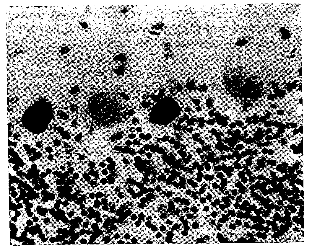
The pathohistological changes in the brain were found to be polymorphic, as were the clinical symptoms, and their diffusion was also established. The morphological changes were distinguished by disorders of the vascular circulation, which were a main constituent of the pathohistological process. In all cases the vascular pathology took the form of perivascular and pericellular oedemas, stasis in the vessels, plasma congestion of the vessel walls, the formation of protein coagulants and vascular dystonia (fig. 1). These symptoms were accompanied by degenerative processes in the nerve cell composition of the brain.
The changes in the cells of the cerebral cortex were distinguished mainly by a pattern of hydropic degeneration and caryocytolysis. The histopathological changes mainly took the form of some kind of degeneration, sometimes in more than one form. Other forms of cell degeneration were infrequent (two cases); they developed either as corrugations or as changes similar to a severe disorder of the nerve cells (Nissl type). Individual nerve cells occasionally showed signs of degeneration in the form of dispersion of the tigroid substance or a change of cell outlines. All types of degenerative process frequently terminated in the death of the cells. This is proved by darkening and the disappearance of cell elements.
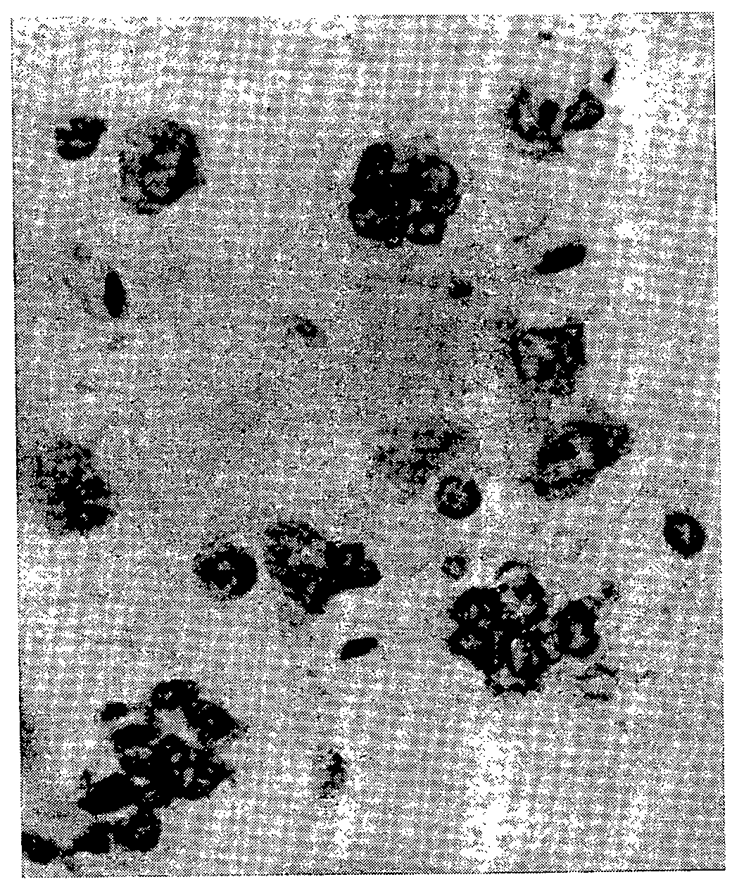
Hydropic degeneration and caryocytolysis were the main symptoms in the subcortical nuclei and in the cortex (fig. 2). Only in infrequent cases were there any other forms of cell element degeneration, in the form of corrugation and severe disorder of the cells (Nissl type) in this part of the brain. Darkening of the subcortical cells was infrequent, but satellitosis was more frequent and more intensive than in the cerebral cortex. Neuronophagia occurred in some subcortical nuclei.
The main symptoms in the medulla oblongata are individual signs of degeneration of the susceptible nerve cells - hyperchromatosis with poorly differentiated nuclei, tigrolysis or dispersion of the Nissl substance, pyknosis of the nuclei and hyperchromatosis of the protoplasm. More clear-cut degenerations as compared with the cortex and the subcortical nuclei are infrequent. There was usually hydropic degeneration of the nerve cells. Corrugation of the nerve cells was observed to result from many forms of nerve cell disorder; it was observed in an experiment after which the dog died. In addition to the pathological processes described, satellitosis and neuronophagia occurred in the nuclei and the medulla oblongata in some cases; darkening was sometimes observed.
The initial stages of degeneration of the cell elements were prevalent in the cerebellum. Hyperchromatosis, often alternating with hypochromatosis, was observed in the purkinje cells. In some cases dispersion of the tigroid substance, with the formation of perinuclear spaces, and a change in the form of the nuclei (fig. 3) were also observed. The initial stages of degenerative processes in the cells were occasionally combined with obvious degeneration in the form of acute swelling of the cells. In isolated cases corrugation of the purkinje cells, cytolosis, and a pathological change in the cells recalling an ischaemic change were noted. In one case in which the animal died after exposure, there were clear signs of neuronophagia of the purkinje cells. Pathological processes which developed in the cerebellum cells frequently led to their death. In this connexion there was a thinning-out of the granular layer and sectoral disappearance of purkinje cells.
The data gained from histological research on acute cannabis resin poisoning show that clearly marked vascular disorders occur in all parts of the brain. Degenerative changes in the parenchyma of the brain were less marked and were also diffused. Animals which died from acute cannabis resin poisoning were found to be suffering from more severe forms of the pathological process. In such cases there was either severe nerve cell disorder (Nissl type) or corrugation of the nerve cells. Other forms of degeneration or individual signs of it were also found, such as hydropic degeneration, hyperchromatosis of the cells, cell shadows etc. There was also pseudoneuronophagia in the ganglion cells and true neuronophagia in the purkinje cells. In these cases and in other experiments where there is acute inhibition of the animal's reaction down to its complete absence, either oedema of the cerebellum or oedema of the medulla oblongata and of the subcortical nuclei is found. The over-all evaluation of histological experiments on acute cannabis resin poisoning is that the pathological process is of the nature of toxic encephalopathy. There is therefore a correlation between clinical developments and the patho-histological process which develops in response to acute cannabis resin poisoning. According to the clinical effects and the morphological changes, the pathological process is encephalopathic in nature.
An analysis of all the electrocardiographic elements of series electrocardiograms recorded on the animals, kymograms and morphological research shows that acute cannabis resin poisoning produces considerable and varied cardiovascular disturbances. The automatism of cardiac contractions is disturbed, the disturbance taking the form of symptoms of marked sinus arrhythmia, sinus tachyarrhythmia or bradyarrhythmia and sometimes of extrasystole. The cardiac contraction rate is disturbed, and the cardiac contractions speed up and slow down at different periods of time. The arterial pressure of animals exposed to cannabis resin poisoning decreases, most of the decrease occurring about 40-90 minutes after exposure.
Electrocardiographic experiments showed changes in nearly all the electrocardiographic elements.
The PQ intervals of most of the animals was found to lengthen. Even during the first few hours of toxic effect a lengthening of the atrioventricular pathway by 0.01 to 0.02 second was noted. An even greater lengthening of the PQ interval, by 0.02 to 0.04 second was noted subsequently during the first and second days. The atrioventricular pathway subsequently returned to normal. These lengthening symptoms point to increased tone of the vagus nerve.
The QRS complex was found to expand as a result of cannabis resin poisoning, increasing by 0.01-0.02 second in most of the animals two hours after exposure, by 0.02 to 0.03 second after 24 hours and by 0.02 to 0.03 second after 48 hours. These changes indicate delayed transmission of impulses by the muscles of the cardiac ventricle.
During different periods the initial correlation between the amplitude of the QT interval and the frequency of cardiac activity was maintained in most of the animals. At the same time a dissociation was noted, in some animals between the QT interval and the frequency of cardiac contractions. Bradycardia was noted at a relatively short QT interval (0.20-0.24 second). At a QT interval of 0.18-0.20 second tachycardia was observed (170-180 per minute). These data indicate that dissociation between the QT interval and the frequency of cardiac activity may occur in cannabis resin poisoning and the lengthening of the QT interval in the case of tachycardia must be evaluated as a sign of impaired metabolism in the myocardium of the ventricles and possibly of degenerative changes.
The T wave was more variable in connexion with the influence of cannabis resin on the animals. Abrupt changes of the T wave occurred, mainly its inversion in combination with a shift of the ST interval. These changes indicate a disturbance of the bioelectric processes in the myocardium in connexion with disturbances of the coronary blood supply. The changes were very marked in all our experiments.
Repeated exposure usually caused increased coronary insufficiency. Disturbances of the cardiovascular system in acute cannabis resin poisoning have a wave-like character, appearing and disappearing. The temporary cessation of symptoms is probably connected with the periodic active mobilization of the compensatory adaptive functions. Nevertheless, in some cases the pathological process developing in the vegetative centres and the cardiovascular system progresses unceasingly and finally leads to derangement of the protective adaptive functions. The electrocardiographic changes and changes in arterial pressure indicate, in our opinion, that pathological processes are occurring in the cardiovascular system, such as various pathological changes in the vessels and a degenerative process in the myocardium. The electrocardiographic and haemodynamic shifts which we observed during the first one or two hours-tachycardia, bradycardia, pronounced sinus arrhythmia, symptoms of coronary insufficiencies and reduced arterial pressure- were the results of damage to the vegetative centres. These changes cannot be explained by the direct toxic effect on the myocardium either clinically or as a long-term effect. The central nervous system-the cerebral cortex, the subcortex, the medulla oblongata with the vegetative centres-and the vascular system of the heart all experience the toxic effect of cannabis resin. This is why the total influence of the poison acts so quickly on changes of the coronary blood circulation and vascular tone. Degenerative changes occur in the myocardium in the course of time due to disturbances of the nervous regulation and to marked vascular disorders, more particularly of the coronary vessels. The pathohistological changes which we found in the central nervous system in the vessels of the heart and myocardium confirm our opinion. In nearly all the animals there were either changes in the cardiac vessels-(plasma impregnation of the vessel walls, stasis in the vessels, diapedetic haemorrhages) or degeneration of the myocardium (cloudy swelling of the sarcoplasm, pyknosis of the muscle nuclei, disappearance of cross striation)-or else all these and others were found simultaneously. Large morphological changes in the brain and in the cardiovascular system do not correlate fully with the degree of markedness of the clinical symptoms, which are less severe than the pathomorphological changes produced in the brain and in the cardiovascular system by acute cannabis resin poisoning. One explanation for this lack of correlation may be the compensatory functions which develop actively during this time.
Changes in respiration were noted as a result of acute cannabis poisoning. As a rule, respiration speeded up within 30 minutes of exposure by an average of 87.5 per cent (41.5±4.32, P0.001, against an initial value of 22.0±1.21). In isolated cases respiration was unchanged after the first 30 minutes. Subsequently, up to 1? hours after exposure almost all the animals still had an increased rate of respiration (32.4±4.84, P0.05; P0.01, against an initial value of 22.0±1.21). During the next two days the respiration rate of individual animals speeded up and in others it fell to below the initial level. The mean values indicated more rapid respiration (P>0.5). On the third day most of the animals had a slower rate (P>0.5). Vascular disorders, such as vascular stasis, congestion or oedema of the alveolar septa and plasma impregnation of the vessel walls, were the main pathological changes in the lungs. In a few cases interstitial pneumonia and a few foci of desquamative bronchopneumonia were observed.
The results of our experiments coincided with the information from other authors ([11] ,[ 12] , [13] , [17] , [18] , [19] , [20] ) that cannabis resin poisoning causes changes in respiration.
Hypersalivation was observed in most of the animals within 30 to 60 minutes after exposure; it usually stopped by the end of the second hour.
For 4 or 5 days after exposure all the animals refused to eat and some of them suffered from diarrhoea and vomiting.
The results of our experiments were in basic agreement with the findings of Chopra R. N. and Chopra G. S. ([11] ), A. K. Strelyukhlin ([12] ,) and Loewe ([13] ,).
However, there is no description by these authors that the animals suffered from anorexia extending to a complete refusal of food.
Morphological changes in the gastrointestinal tract were indicated by various pathological signs, the main one being oedema of the mucous lining and the submucous linings. However, focal infiltration of the mucous and submucous lining, necrotic portions of the surface of the mucous lining and atrophy of the fundus glands of the stomach were rare. Necrosis of the villi, stasis and congestion of the villi, desquamation of the epithelium of the mucous lining, hyperaemia of the muscular layer of the small intestine and degenerative changes of the epithelium of the small intestine glands were rare.
Pathological changes were observed in the liver, pancreas, kidneys, adrenal glands and spleen.
Degenerative changes of the hepatic cells in the form of cloudy swelling were noted in the liver. There were a very few cases of necrotic changes in the cells and of toxic hepatitis.
There were only minor pathological changes in the pancreas and only in a few cases were there degenerative changes in the secretory portion.
Morphological changes in the kidneys in some cases bore over-all resemblances to glomerular nephritis. The glomeruli and tubules had undergone pathological changes. The morphological changes took the form of hypertrophy and mutilation of the glomeruli. Accumulations of serous fluid were found in the cavity of Bowman's capsule, and in some cases desquamated epithelia were found. Various extents of degeneration of the tubules were also noted. In other cases morphological changes were represented in every experiment by two or three pathological symptoms, such as degeneration of the epithelium of the tubules, swelling of Bowman's capsule, necrosis of the epithelium of the distal part of the nephron, congestion of the cortical and medullary layers and focal haemorrhages.
The pathological picture of the adrenal glands varies. Amongst various pathological symptoms the main ones were unequal distribution of lipoids in the cells of the cortical substance and lipoid union of the cortical and reticular zones.
There were considerably fewer pathological changes in the spleen than in other organs and they were distinguished by sinus congestion.
Acute cannabis resin poisoning affects haemopoiesis. On the second day after exposure the red cell count definitely decreased - M=4895000 instead of 6355000 (P0.01). The count then remained at a reduced level for nineteen days inclusive (P0.02; P0.001).
Exposure caused an increase in the leucocyte count, the shift in most cases being noted 30 to 60 minutes after exposure (11.5±3.8 instead of the initial value of 8.6±0.68 thousand in 1 mm[3] ).
There was a definite increase in the leucocyte count on the first and sixth days (19.2±4.96; P0.05; 18.8±2.30 in 1 mm[3] ; P0.001). This was accompanied in some cases by shifts in leucocyte make-up. There was a reduction in the monocyte count within 30 to 60 minutes after exposure, and there was a bigger and more clearly defined decrease on the first and ninth days (P0.01; P0.001). Simultaneously, the lymphocyte counts in most of the animals increased (P0.05; P0.02).
Acute cannabis resin poisoning was accompanied by changes in blood sugar level. The level increased in most animals during the first two hours. The largest and statistically reliable increase occurred 90-120 minutes after exposure (113.5±8.7; P 0.02 instead of the original value of 89.7±2.93 mg). Subsequently the level rose and fell between normal top and bottom limits for nine days. Our experiments with Yujnochuisk cannabis resin showed that the blood sugar level was the same as in the experiments described in the literature with Indian cannabis ([21] ,), mostly varying between the normal top and bottom levels.
A change was observed in the level of adrenalin-like substances in the blood, which rose and fell (10.7±2.117, P0.5; 5.61±1.958; P0.5 instead of 9.713±2.913).
The cholinesterase activity of the animals rose and fell at different periods of time (2.02±0.465, P0.5; 0.93±0.116; P0.5 instead of 1.31±0.286).
The blood plasma protein level rose and fell at different times, the general tendency being to fall. A convincing and statistically reliable drop occurred on the first and
Functional and morphological changes in experimental acute poisioning substances prepared from Yujnochuisk cannabis 7 fifth days after exposure (6.74±0.385; P0.05; 6.02±0.162; P0.001 instead of 7.73±0.279).
Our experiments show that acute poisoning by resinous substances prepared from Yujnochuisk cannabis produces many nervous and somato-vegetative disturbances.
As clinical and morphological research shows, these disturbances arise and develop in association with encephalopathic processes in the brain, with simultaneous disease of the tissues and individual organs.
Yujnochuisk cannabis resin has a wide spectrum of harmful activity and in this connexion is very dangerous to the organism.
(To be concluded)
E. A. Kechatov, Research on resinous extracts of cultivated and wild cannabis growing in the European part of the USSR. Thesis for the degree of Candidate of Pharmaceutical Science, 1961. Summary of Thesis 1962, 1-22.
002Commission on Narcotic Drugs of the Economic and Social Council of the United Nations. E/CN.7/497, 1966, 13-14, 22-24.
003S. Allentuck, K. M. Bowman, The psychiatric aspects of marihuana and intoxication. Amer. J. Psychiatr ., 1942, 99, 248-251.
004A. Woltman, Family and community ideologies. Marihuana problem in the city of New York, Lancaster, 1944, 33-148.
005G. D. Reichard, Marihuana problem. J. Amer. med. Ass ., 1944, 125, 594-595.
006H. B. M. Murphy, The cannabis habit: a review of recent psychiatric literature. Bulletin on Narcotics, 1963, 15, I, 15-23.
007Andrade O. Moraes, The criminogenic action of cannabis (marihuana) and narcotics. Bull. on Narcotics , 1964, 16, 4, 23-25.
008G. Fort, Giver of delight or liberator of sin. Drug use and "addiction" in Asia. Bull. on Narcotics , 1965, 17, 3, 1-11.
009T. H. Mikurija, Marihuana in medicine: past, present and future. Calif. Med., 1969, 110, 34-40.
010V. M. Perelygin, S. H. Altynbekov, Pathomorphology of experimental pneumoconiosis caused by complex organomineral dust in fibre works. Work of the Kirghiz Institute of Epidemiology, Microbiology and Hygiene, 1961, 5, 168-174.
011R. N. Chopra, G. S. Chopra, The present position of hemp
000drug addiction. India. Indian Med. Research Memoirs , 1939, 31, 1-119.
012A. Y. Strelyukhin, Clinic of acute and chronic cannabis resin poisoning. Thesis for the degree of Doctor in Medical Sciences (From the Psychiatric Clinic of the Turkmen Government Medical Institute) 1942.
013S. Loewe, Pharmacological Study. Marihuana problem in the city of New York, Lancaster, 1944, 149-210.
014Cordeiro de Farias, Use of Maconha (Cannabis sativa L.) in Brazil. Bull. on Narcotics, 1955, 7, 2, 5-19.
015E. A. Carlini, C. Kramer, Effects of cannabis sativa (marihuana) on maze performance of the rat. Psychopharmcologia, 1965 (Mar), 7, 175-181.
016S. Garrattini, Effects of cannabis extract on gross behaviour. Hashish, its chemistry and pharmacology, 1965, 70-82.
017P. Mascherpa, M. Bazzi, Wirkung der Cannabis Sativa L. var. indica. Lahm. auf die Atmung. Arch. Exp. Pathol. Pharmacol ., 1941, 197, 306-312.
018G. N. Pershin, The pharmacology of cannabis sativa L. var. indica . "Pharmacology and Toxicology ", 1949, 4, 12, 37-38.
019R. Dagirmanjian, E. S. Boyd, Some pharmacological effects of two tetrahydrocannabinols. J. Pharm. Exp. Therap. ,. 1962, 135, 1, 25-33.
020B. S. Bose, A. Q. Saifi, A. W. Bhagat, Effect of Cannabis Indica on hexobarbital sleeping time and tissue respiration of rat brain. Arch. lnt. Pharm., 1963, 141, 3-4, 520-524.
021C. Miras, Some aspects of cannabis action. Hashish, its chemistry and pharmacology. 1965, 37-53.

