Introduction
Epidemiological study
Methodology
Results
The substances used
Storage, preparation and injection of the substances
Factors conducive to contamination
Current epidemiology of drug addiction in the Paris region
Epidemiology of Candida albicans
Clinical study
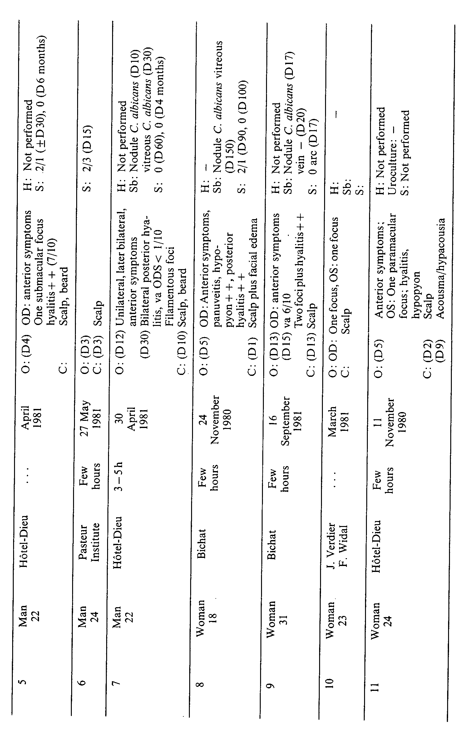
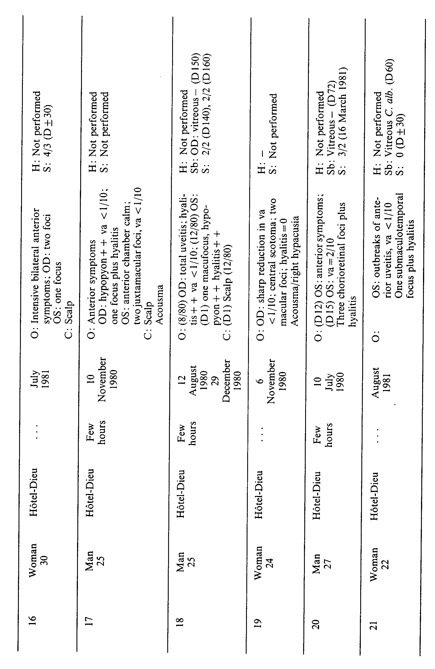
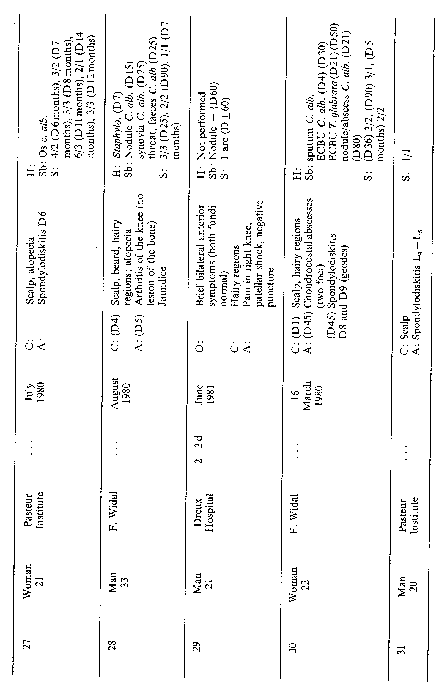
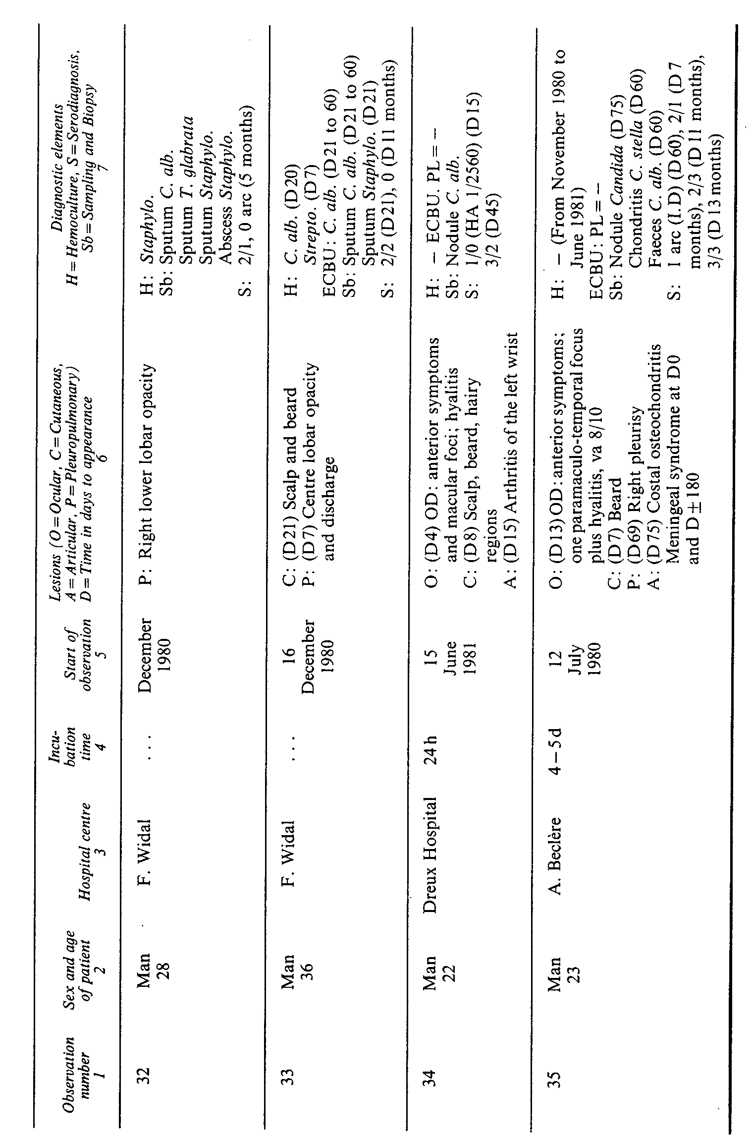
Diagnostic elements
Treatment
Conclusion
Acknowledgment
Author: M. MELLINGER,, O. DE BEAUCHAMP,, C. GALLIEN,, R. INGOLD, M. J. TABOADA
Pages: 61 to 81
Creation Date: 1982/01/01
ABSTRACT
A study of 35 cases of deep-seated Candida albicans candidiasis, affecting heroin addicts in the Paris region, revealed that the lesions observed were mainly cutaneous (88 per cent) and ocular (65 per cent), along with a number of instances of osteoarticular and pleuropulmonary attacks. The clinical and epidemiological findings indicated the possibility that Candida albicans might be transmitted through the heroin. This paper also presents the diagnostic techniques, and the novelty of the clinical picture, in which cutaneous and ocular lesions are frequently associated (57 per cent).
In late November 1980, the Marmottan Medical Centre in Paris was informed that a number of heroin addicts were hospitalized in the ophthalmology department headed by Professor Pouliquen. All of these patients were suffering from severe uveitis whose appearance suggested the possibility of a fungal pathology. The diagnosis was confirmed the following month in other cases, indirectly through serological analysis and by the discovery of Candida albicans as the responsible agent.
Since that time, there has been a steady stream of new cases. While the visceral and particularly the ocular [ 1] types of candidiasis were known, the authors were faced with a new, apparently epidemic phenomenon which has so far never been described, either in France [ 2] or in the United States of America [ 3] .
At the beginning of 1981, the Department of Epidemiology of the Marmottan Medical Centre circulated an information bulletin to the major Paris health-care departments with the aim of co-ordinating a multi-centre research effort on this phenomenon. This effort yielded data on 35 cases. In addition, an epidemiological field study was undertaken on drug addicts not affected by candidiasis and on 20 cases affected by candidiasis.
The purpose of this study was to answer the following two questions: (a) What was the transmitting agent of the Candida albicans? (b) Was the population of heroin addicts particularly exposed to mycoses? It was therefore necessary to take into account the particular characteristics of the addicts and the substances used, and the specific nature of Candida albicans, It will be recalled that Candida albicans is known as a fungus found exclusively on human hosts, a saprophyte of the mucous membranes [ 4] .
The mycological research was conducted mainly by Professor Drouhet of the Mycology Laboratory of the Pasteur Institute (except for observations 3, 11, 17, 19, 26, 29) using pathological samples and sera [from 38 cases (10 of which are not referred to here).]
The field study was carried out by the authors on 20 patients (see annex), three of whom were seen by Dr. S. Dally of the Fernand Widal Hospital.
The research was conducted in stages between November I 980 and October 1981, with no particular selection with regard to the location of the lesions. The patients were interviewed, their consent having previously been obtained, either at the hospital itself or outside any kind of treatment context, in which case care was taken to avoid introducing a therapeutic connotation into the relationship.
The data were collected using a standard form inquiring into about 40 variable factors in such areas as:
The notion of "conducive environment";
The substances used;
The characteristics of the substances most recently used;
The source of supply of these substances;
The method of preparation;
The method of injection;
The equipment used.
In addition, throughout the year, a "field" approach was used which was based on the clinical and epidemiological work of collaborating colleagues at the Marmottan Medical Centre and the Fernand Widal Hospital who supplied general figures on drug addiction in 1981 [ 5] . The addicts themselves, who were not affected by candidiasis, also helped by volunteering samples of substances used.
The first case (observation 30) was observed in March 1980 with 9 further cases observed eight months later (approximately half of them retrospectively). lt is believed that the principal explanation for this increase in number of observations lies in the media information effort undertaken at that time. Subsequently, the frequency of observation appears to have levelled off, with the appearance of an average of 6 new cases per quarter. However, 7 new observations were recently brought to light, bringing the total to 46 observations. It was felt certain that a considerable number of the victims did not seek medical care, since, in 28 per cent of the conversations with patients, confirmation was given of the existence of an analogous symptomatology in persons close to them (spouse, friend, brother etc.), suggesting a candidiasis evolving spontaneously towards an apparent cure, although in fact four of these "pairs" had to be hospitalized (observations 1, 11, 14 and 29).
The population studied consisted of 16 women (from 18 to 31 years of age) and 19 men (from 20 to 35 years of age). In 32 cases the subjects had been taking heroin intravenously for an average of 4 years (from 0 to 14 years). Age and the duration of intravenous addiction were uniformly distributed in the case of both sexes.
Twenty-five per cent of the subjects were using or had used at least four different substances intravenously (e.g. heroin, brown sugar, opium, cocaine, morphine-stimulating substances).
Seventy-five per cent of the subjects were using or had used brown sugar, a variety of brown heroin in granulated form (never in isolation).
Eighty-five per cent were using or had used the white heroin known as "Thai" heroin. This substance was responsible for the intravenous addiction of 83 per cent of the victims of candidiasis presented in this paper.
One hundred per cent had recently used the brown variety of heroinknown as "Iranian" heroin.
This type of heroin, which has been the principal variety available on the market for the past two years, had been used on an average of four months (15 days to 12 months) before the onset of the disease. One case (observation 3) differed from the others in the "recreational use" of opiates; injected on a one-time and isolated basis.
The appearance of this powder was described as "pasty" or even moist (65 per cent), with a colour ranging from light grey to dark brown (75 per cent). It was often difficult to dissolve it in water and it was prepared in lemon juice or in vinegar. In 50 per cent of the cases the injection was accompanied by a less intense "flash" and by associated symptoms such as headaches and vomiting.
Despite the designation "Iranian" given to this substance, its international origin was known with certainty in only 40 per cent of the cases, while its illicit distribution was always made in the Paris region (three patients were residents of Normandy).
Apart from the heroin varieties and auxiliary substances already mentioned, no new practices were involved, such as the licking of the needle (35 per cent) and the use of water from toilets (10 per cent), both potential sources of Candida albicans contamination.
With respect to the practice of injection, pains were taken to determine the percentage of cases in which the subjects injected themselves in one part of the body only (45 per cent), in which abscesses had formed (10 per cent), and in which there was an occurrence of the phenomenon known as "dust" (15 per cent) i. e. the injection, because of poor filtering, of particles (or perhaps transitory bacteremia or a form of anaphylaxis) reproducing a septicemic "mini-syndrome".
With respect to the existence of a condition predisposed to infection specifically mycotic, examination for an external candidiasis, an intercurrent disorder, or some sort of endocrinal imbalance such as diabetes, pregnancy etc., was equally unsuccessful. It should be emphasized at this point that in the six cases in which an external candidiasis was mentioned it occurred after treatment with antibiotics and in a specific connection (bronchial dilation, observation 32; pneumonopathy, observation 30).
It is generally agreed that more and more young people are becoming addicted to heroin at an increasingly early age and that it has become the drug of first resort.
Unfortunately, the use of epidemiology in this area is quite new and by no means readily accepted. Attempts are made to justify this resistance to the epidemiological approach by raising such bugbears as the fear of having one's name on file, loss of anonymity etc., and these have the paradoxical effect of leaving the courts and the police with a monopoly on the unencouraging statistics. For example, as of August 1981 there were in the Paris region almost twice as many drug-related arrests as there were in 1976, and twice as many deaths because of overdose as in 1978, with heroin the preferred substance in 80 per cent of the cases [ 6] .
In this study, the C. albicans species was identified in every case. This predominance of C. albicans is also true of the clinical observation of human beings as a whole, but tends to give way to other species (C. parapsilosis, C. tropicalis etc.) as soon as drug addicts are among the victims of this fungal disorder [ 7] , [ 8] . This applies particularly to cases of endocarditis caused by Candida [ 9] where 46 observations were found, mainly American, during the period 1940 - 1970 in which C. albicans was identified in only 11 per cent of the cases (as opposed to 48 per cent for C. parapsilosis).
The well-known saprophytism of C. albicans would hardly seem to indicate that it might be transmitted through heroin, although the latter is frequently mixed with glucose (nutritive medium) and has a moist con-sistency in 65 per cent of the cases. Experiments to test for resistance to desiccation in this type of medium are currently in progress.
Efforts at direct demonstration were unsuccessfully undertaken, using several small samples. This negative finding was substantiated by the available literature on this subject, where research along these lines has led to positive results in only one instance [ 10] . In the authors' opinion, the failure was a result of improper methodology. Larger samples are required for an analysis of this kind.
There were two characteristic phases: an initial phase marked by a fever syndrome, and a developed phase characterized by the appearance of metastatic lesions.
This phase exhibited all the characteristics of a septicemic syndrome: fever of 39 - 40°C, intense shivering and diffused pain. In one half of the cases observed [ 10] , the lapse of time between the last injection of heroin and the onset of this phase was determined. In 82 per cent of the cases there was a delay of three to eight hours, and in three cases a delay of one to five days. It was interesting to note that in the case of observation 3, this phase was triggered by a single, isolated injection.
Though intense, this septicemic condition is but a weak alarm signal for the drug addict, who is, in fact, familiar with a similar set of symptoms known as "la poussière" ("dust"). This condition, the physiopathology of which is unknown, is believed to come about as the result of the injection of a macroparticle which is allowed to pass because of defective filtering (cotton). Potentially harmful injections of this kind are very often experienced with little anxiety because the symptoms are transitory, lasting some 12 hours at the most, and leaving no after-effects. The condition itself, however, begins shortly after the injection. The fever usually abates within three or four days, but in some 30 per cent of the cases it may last from two to four weeks.
On three occasions (observations 2, 4, 28), this phase was accompanied by an evolutive-jaundice. In one of the patients, a hepatic punch biopsy failed to reveal any fungal element, but as in several other cases, the hepatic tests were complicated by the presence of extraneous factors such as amino-pherase and alkaline phosphatase enzymes. These modifications were difficult to interpret since they occurred frequently in the cases of opiate drug addicts [ 11] , just as did the signs of viral hepatitis.
Five observations indicated an otorhinolaryngological symptoma-tology which was totally inexplicable and which consisted of unilateral or bilateral acousma, sometimes complicated by hypacusia, especially in the low-frequency region. These symptoms disappeared of their own accord within three or four weeks. On the one hand it is difficult to ascribe them to the Candida. On the other, one is inclined to wonder whether the lemon juice used might not have had a toxic effect on the ear, since this kind of symptom among opiate drug addicts was never noted before.
The metastatic lesions appeared in association with one another (see figure). Among the 35 cases observed, four principal loci could be distinguished:
Cutaneous lesions (88 per cent);
Ocular lesions (65 per cent);
Osteoarticular lesions (17 per cent);
Pleuropulmonary lesions (8 per cent).
The typical pattern involved an association of cutaneous and ocular lesions (57 per cent). In two cases (observations 34, 35), more complex patterns combining three and four loci were observed.

The lesions appeared on an average of 7 days ranging from 1 to 21 days after the initial phase. Developing in successive stages, they often manifested themselves in the form of quite large papulo-nodular sores (1 - 3 cm in diameter) which were occasionally painful when located in the scalp (90 per cent), at which time they became noticeable. More frequent in men, the lesions extended to the beard and the hairy regions of the body (under the arms, in the pubic region, and on the chest and limbs). They were accompanied by satellite adenopathies. They also appeared in the form of quite ordinary pustules.
Histologically speaking, biopsies of the nodules revealed a polymorphous infiltrate with more or less degraded polynuclear cells at and around the hair follicle. Staining with para-aminosalicylic acid revealed mycelial filaments and yeasts in the hair and the perifollicular infiltrate - one of the aspects of Candida-caused acute folliculitis [ 12] . Candida was also obtained by sampling from the surface of the lesions.
In three cases, this pustular folliculitis evolved into a superacute condition, developing in one instance (observation 30) within 48 hours into a purulent spot on the scalp, or being accompanied by intense reaction signs, edematic infiltration of the face and desquamation of the palms and soles of the feet.
In the majority of cases, however, the clinical picture was subacute and the condition could even go unobserved.
In four cases (observations 22, 23, 24, 25) the cutaneous lesions occurred in isolation. Nevertheless, as in the cases of uveitis, a number of symptoms which occurred at the same time as or slightly before the skin eruption, were found in the anterior chamber of the eye (photophobia, conjunctival hyperemia etc.) but passed quickly, leaving no after-effects.
In all of the cases the conditions improved spontaneously until they disappeared altogether, leaving no sequelae. When treated, the lesions stabilized within three weeks, although some fresh outbreaks were observed under treatment (observations 9,17). Where they were left untreated, the outcome was just as favourable, as indicated by the many cases where there was no hospitalization as well as by observation 35, where there was spontaneous sterilization of the lesions in six to eight weeks.
The ocular lesions accounted for the seriousness of the clinical picture. The symptomatology was that of an infectious uveitis which, although extensively described in available literature [ 13] on this subject, had never been encountered before on such a wide scale. Seventeen cases affected one eye and six affected both eyes. In terms of locus and degree of intensity, this symptomatology could be divided into two forms [ 14] .
1. Localized forms (26 per cent)
This form was a predominantly posterior uveitis in which the retinal or retino-vitreous foci of the disorder, frequently more than one (two to four foci), exhibited a poorly delimited white-yellowish appearance. In general, no papillary edema was observed.
2. Diffuse forms ( 74 per cent)
These forms were accompanied by intense and diffuse inflammation which could develop into full-scale endophtalmitis accompanied by pink-eye and photophobia. The anterior chamber of the eye was the seat of a cellular reaction with dense Tyndall phenomenon, leading in the worst cases to a hypopyon. Often, the victim's visual acuity could scarcely be measured, being limited to the mere perception of light. The vitreous also is the seat of a severe hyalitis, which could mask the back portion of the eye. Whitish, poorly delimited foci could similarly occur around the posterior pole. Associated symptoms frequently included pronounced vascularity and papillary edema. In two cases (observations 9, 34), the veins contiguous to the foci were affected by a thrombophlebitic condition.
On the average, the ocular lesions occurred within 8 days (2 - 15 days) in the case of the anterior chamber symptoms and 15 days (3 - 30 days) in cases involving an impairment of visual acuity.
The evolution of the lesions was that of a non-specific sequela pathology.
1. Vitreoretinal disorder
The vitreous organization secondary to the infection and inflammation syndrome took the form of more or less retractile adhesions (observation 13), which in extreme cases could lead to a massive retraction of the vitreous membranes, possibly causing a total, complex, and fixed detachment of the retina.
2. Chorioretinal scars
The cicatrization process in this case will proceed atrophically, causing the necrosis of the internal layers of the retina and the choroid (observation 18), or hypertrophically giving rise to a fibroproliferative organization (observation 1), often neovascular and hemorrhagic in character. These lesions result in scotomata at various sites: central if the scar is macular, and paracentral (observation 21) when the lesion is papillomacular.
3. Other sequelae
These are possible and may take a very advanced form: cataracts or atrophy of the ocular globe (observation 8).
In 21 observed cases the lesions were found to evolve as follows:
In 48 per cent, stabilization of the foci after treatment (period variable);
In 19 per cent, healing with total recovery of the back of the eye (observation 35, spontaneous healing in two months);
In 33 per cent, serious after-effects (total loss of vision or acuity of 3/10°).
Apart from antifungal chemotherapy, topical treatment was important - laterobulbar injections of corticosteroids, evacuative puncturing of the anterior chamber, and above all, vitrectomy.
The indications were determined as a function of the initial attack, the macular topography of the foci, the inefficaciousness of the chemotherapy, and evolution in the direction of retraction.
A total of eight vitrectomies were tried (observations 7, 8, 13, 14, 15, 18, 20, 21). Of these, three produced good results, while the remaining five were performed on patients suffering from advanced disorders (retinal necrosis) and were complicated in three cases by retinal detachment.
These were found under eight observations classified into three distinct forms:
l. Costal osteochondritis (observations 26, 30, 35)
These lesions were quite specific in appearance and involved an extensive exaggeration of the convexity of the thorax in the area of the costal arches or the sternocostal junction. They took the form of genuine parietal abscesses requiring reduction by surgical means. Anatomopathological examination indicated the different planes of the wall affected by the polymyositis, chondritis or osteitis (observation 35). The lesions were accompanied by fever, their appearance being preceded by severe pain. Each of the swellings followed its own course and disappeared spontaneously (observation 30).
2. Spondylodiskitis (observations 27, 30, 31)
The warning symptoms may be so mild that the physician is consulted only at a late stage (observation 27), or they may be sufficiently severe to suggest an acute infection. Bone scintiscans show hyperfixation, while radiotomography reveals osteolytic images of the vertebral bodies (observation 30). The prognosis under treatment is favourable, though with a readjustment of the intervertebral space.
3. Nonoarthritis (observations 28, 29, 34)
The lesions were abarticular and did not attack the bone. They appeared early and their evolution was characterized by variable swelling and redness around the joint. The wrist was affected on one occasion, the knee on two occasions.
l. Bronchopneumonic disorders (observations 32, 33)
The systematically encountered foci (the lobus medius and the apical segment of the lobus inferior dexter) resisted treatment on the basis of antibiotics and evolved over a long period (three to five months) to the accompaniment of fever. Candida albicans was never discovered by itself, but in the company of Staphylococcus pyogenes or other saprophytes (Torulopsis glabrata). Despite treatment, a lengthy period was required before it disappeared altogether from the patient's sputum.
2. Pleurisy disorders (observations 33, 35)
These were always of the inflammatory type, either in reaction to a pulmonary focus (observation 33) or apparently in isolation.
In concluding this section, particular attention is drawn to the specific association of the cutaneous and ocular lesions - with the latter predominating by far in the clinical picture - and to the possibility, in the case of most of the lesions and particularly the cutaneous ones, of spontaneous evolution towards apparent recovery.
In this connection, it should be noted that observation 35 was quite exceptional: there was evidence suggesting a meningeal attack (lumbar puncture = 300 elements) accompanying a combination of four associated lesion foci: ocular, cutaneous, pleural, and osteochondral. After 12 months, during which the condition was allowed to develop spontaneously, the patient was re-examined and found to be free of any detectable after-effects, although the serological picture remained positive (a report is to be prepared by Dr. J. F, Delfraissy, Antoine Béclère Hospital).
The aetiological diagnosis is based essentially on the findings of the mycological and serological analyses.
This diagnosis was made on the basis of several types of pathological samples:
Cutaneous samples: The cutaneous samples were obtained either by scraping the surface lesions or by biopsy of the folliculo-nodular elements. They were then directly examined and placed in a Sabourand culture medium.
In 75 per cent of the cases fungal elements were found to be present.
Ocular samples: The conjunctival smears and punctures of the anterior chamber were in every instance negative. On the other hand, of the eight vitrectomies carried out, three vitreous cultures developed. Several authors have already called attention to the difficulty of substantiating the presence of Candida albicans in this medium [ 1] , [ 13] .
Other tissue samples: Candida albicans Was also discovered in the osteoarticular lesions (synovial, os, chondrocostal abscesses) as well as in the customary places (throat, sputum, stool etc.).
Hemocultures: Unfortunately, hemocultures were prepared in only 37 per cent of the cases observed, with the findings positive in only two instances. It is worthwhile recalling in this connection that candidemia is an intermittent phenomenon [ 15] because of the filtering role placed by certain organs (liver, spleen, ganglions etc.). A number of writers go so far as to recommend that hemocultures be taken from various points of the vascular bed. In cases of Candida endocarditis there is a 15 per cent rate of false negative findings [ 9] .
It should be noted at this point that a uroculture has a diagnostic value similar to that of a hemoculture.
It was also possible through laboratory analysis to reveal the mixed character of certain of these deep candidiases: three hemocultures developed for streptococcus or staphylococcus as well as three cultures of abscessed veinitis containing no Candida.
Candida albicans was identified by the filamentation test and the observation of chlamydospores. Mycogrammes failed to reveal any particular resistance to antifungal agents.
A total of 28 observations were carried out using the electrosyneresis method, which remains the preferred technique because of its reliability and speed, these being such that the evolution of the precipitins over time could be easily observed [ 16] , [ 17] .
Of the serological tests performed, 75 per cent revealed two to six precipitin arcs, a rate which suggested the presence of a deep candidiasis. It should be emphasized, however, that serodiagnosis must be conducted using several samples and analysed on a case-by-case basis. In general, the findings became negative within three to four months.
Virtually no tests for cellular immunity were carried out. In one case a high (immunoglobulin E) level was found, and in another an intense and immediate reaction to candidine.
The principal chemotherapeutic agents employed are simply listed, since it is too early to be able to assess the specific efficacy of the antimycotic substances used.
Flucytosine (5 FC) (proprietary name Ancotil), used at a daily dosage of 200 mg/kg;
Amphotericine B (proprietary name Fungysone), administered intravenously at a dosage of 0.1 - 1 mg/kg, with monitoring of the kidney function;
Ketoconazole (not commercially available), at a dosage of 400 - 600 mg/day.
It should also be noted, however, that the more severe conditions were treated with amphotericine B administered intravenously either alone or in association with 5 FC, the aim being to obtain synergy in a cumulative dose of nearly 1 g, a level seldom reached because of the nephrotoxicity or the treatment or the awkwardness of its administration. By combining it with ketoconazole or 5 FC, a cumulative effect was achieved.
No general conclusions could be drawn. At best, a few factual observations and hypotheses can be offered:
All the evidence suggests a pathology, epidemic in behaviour, whose origins can only approximately be traced back two years and whose significance in quantitative terms can be estimated only imperfectly;
There have been no noticeable changes in the circulation and use of the narcotic substances, which would tend to argue against the explanation that the subjects themselves or the group is the contaminating agent;
The same type of heroin, brown but of uncertain international origin, was used by all the persons affected. This product must therefore be regarded as a possible transmitting agent;
If one accepts this mode of transmission, the ability of Candida albicans to survive in this medium must be reconsidered. The negative findings obtained with small heroin samples do not represent a valid counterargument, since it is the authors' belief that the methodology was inadequate.
Finally, on the basis of the clinical picture (and not the lesions per se), the possibility of a particular immune area in drug addicts and a new pathogenesis for the C. albicans species found should be considered.
Extended study alone should be able to provide the answers to these questions.
Thanks are due to the following for their close co-operation in the study: Dr. Dally, Dr. Bartholomy, Dr. Delfraissy, Dr. Di Constanzo, Dr. Drouhet, Dr. Dupont, Dr. Mallecourt, Dr. Mechali, Dr. Péan, Dr. Thierman, Dr. Cole, Dr. Leandri, Dr. Castel, and the departments under the direction of Dr. Pouliquen, Dr. Bastin, Dr. Herreman, Dr. Lapresles, Dr. Rousselie, and Dr. Drouhet.
R.A.Getnick and M.M. Rodrigues, "Endogenous fungal endophthalmitis in drug addicts", American Journal of Ophthalmology , vol.77, No.5 (1974), pp. 680-683.
002S.Dally and G. Thomas, "Candidoses chez des toxicomanes", Nouvelle Presse Médicale , vol.10, No.23 (23 May 1981), p.1941.
003B.Cole,New York Bellevue Hospital, personal communication, June 1981.
004E.Drouhet,"Champignons opportunistes et mycoses iatrogènes", Bulletin de l'Institut Pasteur , vol.70, 1972, pp. 391-464.
005R.Ingold and others, "Epidémiologie de la pharmacodépendence", Bulletin du Department d 'Epidémiologie de Marmottan/Institut National des sciences et de la Recherche Médicale, Avancement des travaux , October 1981.
006P. Benquet, "La brigade des 'stups' face à laremontée de la drogue", Le Monde , October 1981.
007C.E. Cherubin, "The medical sequelae of narcotic addiction", Annals of Internal Medicine , vol.67, 1976, p. 23.
008D.B. Louria, T. Hensle and J. Rose, "The major medical complications of heroin addiction", Annals of Internal Medicine , vol. 1, 1967,p. 1-22.
009Mellinger, "Les endocardites à Candida: analyses de 200 cas", Doctoral dissertation (Paris, Université R. Descartes, 1978).
010Joachim and S.H. Polayes, "Subacute endocarditis and systemic mycosis", Journal of the American Medical Association, vol. 115, 1940, pp. 205-208.
011Dally, J. Dugarin and E. Fournier, "Le foie des toxicomanes", Concours Médical , vol. 102, No. 30 (July 1980), pp. 4311-4314.
012Drouhet and others, "Nouvelle pathologie cutanée: candidose folliculaire et nodulaire au cours de septicémies à Candida albicans chez des héroinomanes", Journées dermatologiques de Paris , 14 March 1981.
013L. AguiIar and others, "Candida endophthalmitis after intravenous drug abuse", Archives of Ophthalmology , vol. 97, 1979, pp. 96-100.
014Communication from Dr. F. Bartholomy of Professor Y. Pouliquen's Department, Hotel-Dieu.
015H.Stone, "La septicémie à Candida", Annals of Surgery , vol. 179, No.5 (1974), pp.1 and 35.
016Drouhet, "Eléments de diagnostic des mycoses profondes", Chemioterapia Antimicrobia , vol. II, No.3 (1979).
017E. Drouhet and B. Dupont, "Acquisitions récentes et perspectives d'avenir dans le traitement des affections à Candida", Revue Francaise d 'Allergologie , vol.16, No.5(1976),pp.291-295.